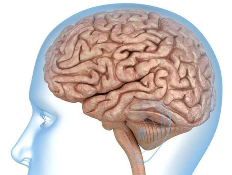
The foramen rotundum is an aperture of circular shape that links the pterygopalatine fossa with the middle cranial fossa. It is found in the medial and anterior part of the sphenoid bone, to be more precise, in its greater wing.
This aperture gives passage to the maxillary nerve. This nerve travels in the skull by the pterygopalatine fossa and the foramen rotundum and exits the cranium through them.
The foramen rotundum belongs to the many circular foramina or apertures that are located in the anterior and medial part of the sphenoid bone. These structures are at the base of the skull.
In this article, you can find information about the foramen rotundum, its structure, its location, and its functions.
Table of Contents
What Are Foramina?
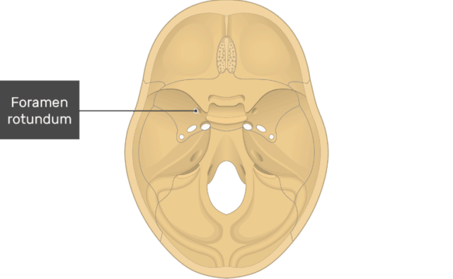
A foramen is a bony aperture in the skeletal structure. Its function is to allow the passage of nerves, veins, arteries, ligaments, and muscles through them.The plural form of foramen is foramina.
There are several small and big foramina located at the base of the skull that serves as pathways of numerous structures as enumerated below.
These are the major foramina found in the cranial base:
a. Foramen Magnum
It makes way for the membrane tectoria, the apical ligament of dens, the spinal accessory nerve, the vertebral arteries, and the medulla oblongata (spinal cord)
b. Mandibular Foramen
It lets the inferior alveolar nerve, inferior alveolar artery, and the inferior alveolar vein pass through.
c. Mental Foramen
The mental vein, artery, and nerve travel through it.
d. Parietal Foramen
It lets the emissary vein from the superior sagittal sinus go through it.
e. Mastoid Foramen
It provides the path for the meningeal branch of occipital artery and the emissary vein to sigmoid sinus.
f. Foramen Cecum
The emissary vein travels through it.
g. Greater Palatine Foramen
It allows the passage of the greater palatine nerve, the greater palatine vein, and the greater palatine artery.
h. Lesser Palatine Foramen
It lets the lesser palatine vein, the lesser palatine artery, and the middle and posterior palatine veins pass through.
i. Incisive Canal
It makes room for the passage of the greater palatine vessels and the nasopalatine nerve.
j. Supraorbital Foramen
It lets the supraorbital vein, the supraorbital artery, and the supraorbital nerve passes through.
k. Superior Orbital Fissure
It makes way for the superior ophthalmic vein, the trigeminal nerve (V3), and abducens nerve, the trochlear nerve, and the oculomotor nerve.
l. Inferior Orbital Fissure
It gives passage to the infraorbital vessels, the zygomatic nerve, and the anterior branches of the sphenopalatine ganglion.
m. Foramen Rotundum
It makes way for the maxillary nerve (V2).
n. Foramen Ovale
It provides passage to the mandibular nerve (V3), the lesser petrosal nerve, and the accessory meningeal artery.
o. Foramen Spinosum
It provides passage to the internal carotid artery with venous and sympathetic plexus, the meningeal branch of the ascending pharyngeal artery, and the greater and deep petrosal nerves from the nerve of the pterygoid canal.
p. Stylomastoid Foramen
The facial nerve and the posterior auricular artery (stylomastoid branch) pass through.
q. Zygomaticotemporal Foramen
It gives passage to the zygomaticotemporal nerve.
In this article, we will focus on the foramen rotundum and its characteristics.
Description Of The Foramen Rotundum
The foramen rotundum is part of the foramina that are present in the sphenoid bone in its medial and front section. This bone located at the base of the cranium. It is one of the foramina or circular openings that are located in this zone.
Etymology Of Foramen Rotundum
The name of the foramen rotundum originates from the Latin language. It comprises from two words, that is, “foramen” and “rotundum.”
The foramen is a Latin word that has the meaning of “hole like an opening,” perforation, or aperture. It is derived from the Latin word “forare” which means to bore or perforate.
Rotundum is a descriptive Latin word that means round.
Hence, we can say that foramen rotundum means “round hole-like opening,” which describes the circular or round shape of this structure of the skull.
Location & Structure Of Foramen Rotundum
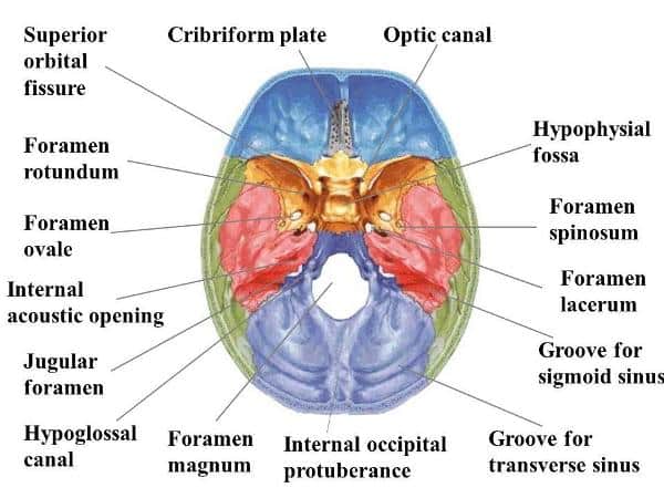
This foramen, in particular, is in between the inferior medial and the superior orbital fissure at the base of the sphenoid, in its greater wing. The lateral wall of the sphenoid sinus forms its medial border.
The whole structure is located in the middle cranial fossa. The foramen goes laterally downwards following an oblique line to join the middle cranial fossa and the pterygopalatine fossa.
The pterygopalatine fossa is a depression in the cranium. It is connected with the oral and nasal cavities, the middle cranial fossa, the infratemporal fossa, the orbits, and the pharynx through eight different foramina of the skull.
There are two pterygopalatine fossae in the human skull, one on the left side, and the other one the right side. They are cone-shaped and located deep inside the infratemporal fossa. They are found on the posterior side of the maxilla on either side of the cranium.
The middle cranial fossa has the function of supporting the pituitary gland. It is formed by the sphenoid bone in its central part, and it has a bony eminence known as the sellaturcica, which is of saddle shape.
The pituitary gland is contained in the central portion of the middle cranial fossa while its lateral portions accommodate the brain’s temporal lobes.
The lateral depressed parts of this fossa are formed by the sphenoid bone’s greater wings as well as the squamous and petrous parts of the temporal bones.
Hence, the fossa has a total of three bones, which are the sphenoid and the two temporal bones. The fossa is filled with many bony landmarks.
The foramen rotundum is circular and occupies a considerable small mean area. This indicates that it does not have a highly significant role related to the dynamics of the venous system of the human head or the blood circulation.
However, the foramen makes way for the maxillary nerve branch of the trigeminal nerve, for the artery of the foramen rotundum, and for the emissary veins to pass through.
Development Of Foramen Rotundum
The foramen rotundum is known to be a circular aperture located in the sphenoid bone to connect the pterygopalatine fossa and the middle cranial fossa.
The development of the structure of the foramen rotundum starts in the fetal stage, and it continues from birth to adolescence. The foramen undergoes structural evolution continuously throughout the process of fetus development.
A ring shape of the foramen is acquired after the 4th fetal month only. The ring is slightly oval in outline during the fetal period, and it achieves a well-defined circular shape after birth.
The diameter of the foramen rotundum is approximately 2.5 millimeters in just born babies, and it can grow up to 3 millimeters by the age of fifteen to seventeen. The average length of diameter in adults is found to be 3.55 millimeters.
Function Of Foramen Rotundum
The foramen rotundum is situated at the greater wing’s anterior base of the sphenoid bone. It is located in the skull.
The foramen transmits or gives passage to the maxillary branch (V2) of the trigeminal nerve (CN V).
The nerve exits from the cranium via the foramen rotundum and the pterygopalatine fossa to travel to the facial zone.
The Maxillary Nerve
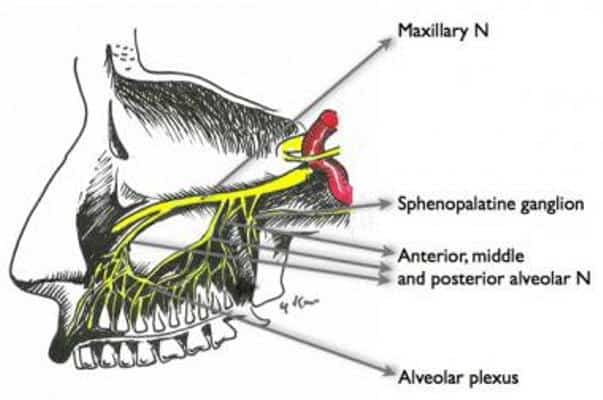
The importance of the foramen rotundum lies in its function to make way for the maxillary nerve from the skull to the facial zone.
The maxillary nerve (CN V2) is one of the three branches that belong to the trigeminal nerve, which is the fifth nerve of the cranium, therefore represented by “V.”
The maxillary nerve has the function of transmitting sensory fibers from the nasal cavity, the sinuses, the maxillary teeth, and the skin that is found between the palpebral fissure and the mouth. It provides sensation or innervates the maxillary, nasal cavity, palates, sinuses, and midface overall.
The maxillary nerve is said to be an intermediate between the mandibular nerve and the ophthalmic nerve in size and position.
This nerve originates in the shape of a flattened band in the center of the trigeminal ganglion in the cranium. It moves forward horizontally and passes through the foramen rotundum to exit the skull after acquiring a cylindrical form and more rigid texture.
It continues its path by crossing the pterygopalatine fossa, and it inclines on the back of the maxilla, entering the orbit through the lower orbital fissure.
After going forward through the orbit’s floor, the nerve reaches the face through the infraorbital foramen. Once there, it divides into the lateral nasal, palpebral and labial branches.
The infraorbital branch of a part of the maxillary artery and the corresponding vein accompany this nerve in its path.
Branches Of Maxillary Nerve
The branches of the maxillary nerve can be divided into four groups according to the location, as follows:
- The cranium: It accommodates the middle meningeal nerve.
- From the pterygopalatine fossa originate the zygomatic nerve, the nasopalatine, the greater and lesser palatine nerves, the posterior superior alveolar nerve, and the pharyngeal nerve.
- The infraorbital canal: It contains the infraorbital nerve, the anterior superior alveolar nerve, and the middle superior alveolar nerve.
- The face: It shelters the superior labial nerve and the inferior palpebral nerve.
The function of the maxillary nerve includes supplying the face with its cutaneous branches as well as carrying postganglionic fibers and parasympathetic preganglionic fibers from and to the pterygopalatine ganglion.
Trigeminal Neuralgia
Damage in the maxillary nerve is associated with pain in the cheekbone, nose, upper lip, and upper teeth. These symptoms take place due to a condition is known as trigeminal neuralgia, which is a facial nerve disorder.
Symptoms
The accompanying symptoms include:
- Intermittent severe pain in the affected region.
- Difficulty in routine activities, including sleeping and eating.
- Sleep and food deprivation due to the fear of unpredictable pain attacks. This can turn into malnutrition.
- Anxiety, depression, and irritability.
- Spread of the pain to the mandibular nerve, thus affecting the jaw, lower cheek, and lower lip.
- The onset of characteristic muscle spasms accompanying the pain. This has led to colloquially naming trigeminal neuralgia as “tic douloureux.”
Causes
The causes of trigeminal neuralgia are not accurately determined.
However, the following factors can lead to the disorder:
i. Compression of the trigeminal nerve by the nearby blood vessels, the aneurysm, or tumor growths.
ii. Inflammation triggered by Lyme disease, sarcoidosis, and multiple sclerosis.
iii. The presence of collagen vascular diseases like systemic lupus erythematosus and scleroderma.
The treatment of trigeminal neuralgia often consists of medications, radiotherapy, and even surgery in severe cases.
It includes:
- Anticonvulsant medication like Tegretol (carbamazepine) is usually prescribed for idiopathic trigeminal neuralgia.
- Second line drugs apart from carbamazepine that includes Gabapentin (Gabarone, Neurontin), phenytoin (Dilantin), and baclofen. They are used to control the pain along with carbamazepine in cases in which carbamazepine loses its effect with time.
- When multiple sclerosis patients develop trigeminal neuralgia, Lamotrigine (Lamictal) is often prescribed for them.
- Radiation therapy and surgery can also become treatment options when the pain is immune to the recommended medications.
Narrowing Of Foramina
Foramina are apertures in the skeleton that serve as pathways for the passage of structures such as blood vessels, muscles, or nerves.
The foramina can narrow in size due to the displacement of the surrounding tissue. This, in turn, restricts the space for the spinal nerves to pass through, and it can result in severe damage and pain.
This condition usually occurs only in the foramina that are in the vertebrae of the spine.

